

Imaging
CT (CAT) Scanning
In a nutshell:
CT or CAT scans combine a series of X-ray images taken from different angles to create cross-sectional images – or “slices” – of the bones, blood vessels and soft tissues inside your pet’s body. These images provide more detailed information than plain X-rays.
The clinical version:
Computed Axial Tomography (CAT), also known as Computed Tomography (CT), is a form of diagnostic imaging that uses X-rays, X-ray detectors and computer processors to create digital images of the scanned area of the patient.
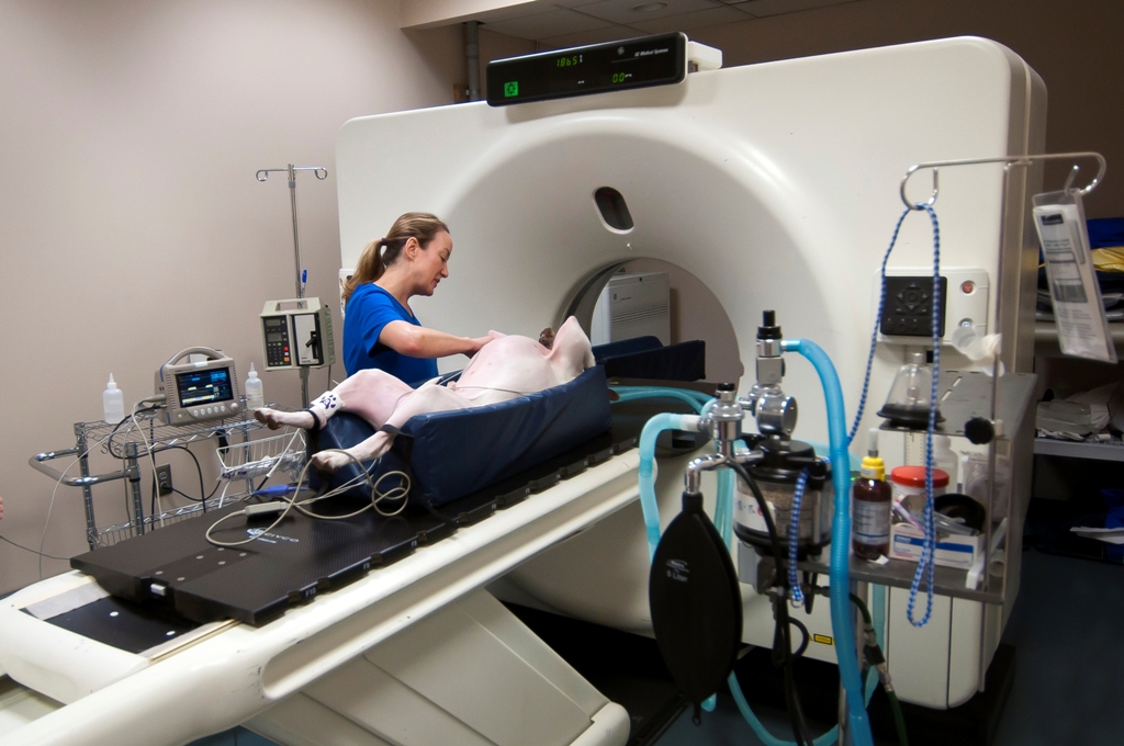
During a CT scan, the patient is positioned on a table that moves the pet through a ring containing the x-ray source and the X-ray detectors. The CT images are cross-sectional “slices,” as if the patient was cut like a loaf of bread, creating dozens to hundreds of digital images. These slices can be examined one by one to reveal the details inside and help identify abnormalities. Contrast agents containing iodine are administered intravenously as part of the scanning process to enhance visualization of abnormal soft tissues and blood vessels.
PetCure Oncology at VRIC uses a 32-slice helical CT scanner to rapidly acquire images of dogs, cats or other pets. The image acquisition itself generally takes less than a minute. A short general anesthesia is necessary so the patient remains in the same position for the duration of the scan, allowing for optimal image quality and enhanced patient safety. Additionally, further diagnostic testing can often be performed while the pet is still under anesthesia, such as a biopsy or fine needle aspirate. These additional tests may be recommended as a result of the CT image finding(s) and/or at the request of your referring veterinarian.
The entire duration of a CT appointment from admission to discharge can vary between 40-90 minutes depending on the size of the patient, complexity of the exam, number of body regions examined, and the indication for further diagnostic testing or procedure such as biopsy, fine needle aspirate or chest tap.
After the CT scan is acquired, CT images can be processed and reconstructed into two- and three-dimensional images for further analysis by our board-certified radiologists.
Our equipment:
Our equipment includes a GE LightSpeed VCT 32-slice helical CT scanner, one of the fastest and best available in the region.
MRI Scanning
In a nutshell:
MRI uses a strong magnetic field and radio waves to create detailed images of organs and tissues in your pet’s body. The MRI machine produces detailed, high-contrast images of bone and soft tissue structures of the brain, spine and joints from any angle.
The clinical version:
Magnetic Resonance Imaging (MRI) uses a very strong magnetic field to align the natural spinning of water molecules within body tissues. MR images are formed by tiny radiofrequency signals generated as the nuclei spin. The molecular alignment that occurs on the sub-microscopic level cannot be felt and has no known harmful effects. The radio signals are collected by small antennae called receiving coils, which are placed outside the patient near the area we are interested in evaluating. An advantage of MRI is its ability to produce images that are simple cross-sections as well as from any other angle with equal resolution. MRI scans give the best soft tissue contrast of all the imaging modalities.
Our equipment:
Our equipment includes a 1.5 Tesla GE Signa Advantage MRI, which is one of the highest field strength MRI units available for animals in the state of New Jersey.
Ultrasound
Ultrasound uses high-frequency sound waves to capture images of the internal structure of soft tissues, especially in the abdomen, chest and neck. It is often used in conjunction with CT as a safe and effective way to assist in reaching a definitive diagnosis. All ultrasounds conducted at PetCure Oncology at VRIC are performed by a board-certified radiologist. Results are relayed to the referring veterinarian the same day the ultrasound is performed.
Equipment Good Enough for People in a Safe Environment Designed for Pets
PetCure Oncology at VRIC affords pet owners access to advanced cross-sectional imaging for their pets on an outpatient basis in an environment specifically designed to minimize risks to their four-legged family members.
We optimize patient safety by using specialized, MRI-compatible patient monitoring and anesthesia delivery equipment in the MRI suite. Similar advanced patient monitoring equipment is available for patients under anesthesia for CT and radiosurgery (if necessary). We use Sevoflurane gas for rapid recovery from anesthesia. We evaluate patient heart rate, breathing rate, blood pressure, oxygen level, exhaled gas and electrocardiogram activity during the scans so potential complications are identified immediately and anesthetic risks are minimized.
The imaging equipment is all manufactured by GE and maintained in top working condition under a service contract with Oxford Instruments OiS technicians, who also provide service in many human imaging facilities. By using equipment manufactured and maintained by highly reputable service providers, we ensure that our patients benefit from the best technology available.
Image Interpretation
A board-certified veterinary radiologist is available at the center to assist in determining the imaging modality of choice and to provide the most accurate image interpretation possible. We do our best to minimize the time a patient spends away from their owners during a treatment or scanning procedure. In most cases, the pet is back with their owner before all of the images have been reviewed and a final radiologist report is generated. Written results of the imaging studies are provided to the referring veterinarian managing the patient's care within 24-48 hours of the imaging study. A digital copy of the CT or MRI is provided to the owner at the time they pick up their pet. Digital copies of the studies are transmitted to referring veterinarians and off-site hospitals when requested.